Get Complete Project Material File(s) Now! »
Triacylglycerol Synthesis
Triacylglycerols (TAG) consist of three fatty acid chains esterified to a glycerol backbone. Triacylglycerol synthesis is an efficient form to store fatty acids and play an essential role in energy storage and energy balance in eukaryotic cells (Ageitos et al. , 2011; Athenstaedt and Daum, 2006; Rajakumari et al., 2008; Sorger and Daum, 2003).
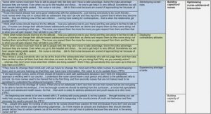
TAG synthesis generally follows the Kennedy pathway which occurs in the endoplasmic reticulum (ER) and requires acyl-CoA and glycerol-3-phosphate (G3P). It is reported that G3P can be produced either from glycerol via glycerol kinase (GUT1) in the cytosol or can be synthesized from dihydroxyacetone phosphate (DHAP) by G3P -dehydrogenase (GPD1) which is a reversible reaction catalyzed by an isoform of G3P dehydrogenase (GUT2) (Beopoulos et al., 2009a; Rossi et al., 2011) (Figure 1. 3). Deletion of GUT2 gene redirected carbon flux toward TAG assembly, with a 3-fold increase in lipid accumulation (Beopoulos et al., 2008).
The first step of TAG biosynthesis starts with the acylation of G3P by the G3P – acyltransferase (SCT1) to form lysophosphatidic acid (LPA) at the sn-1 position. LPA can also be formed by reduction of acyl-DHAP by an NADPH-dependent acyl-DHAP reductase (AYR1) (Beopoulos et al., 2011; Rossi et al., 2010).
The further acylation of LPA is catalyzed by LPA-acyltransferase (SLC1) to generate phosphatidic acid (PA) at the sn-2 position. This is followed by PA dephosphorylation via the PA-phosphohydrolase (PAP) which releases the diacylglycerol (DAG) (Beopoulos et al., 2009a).
In the last step of de novo synthesis of TAG, the synthesized DAGs can follow different ways, either by using diverse acyl donors, acyl-CoA dependent such as DGA1 with acyl-CoA as an acyl donor or with acyl-CoA independent phospholipid:DAG acyltransferase (PDAT) e.g. LRO1 with glycerol phospholipids (PL) as an acyl donor (Dulermo and Nicaud, 2011).
Ex-novo Lipid Synthesis
The lipid accumulation performed from hydrophobic substances (such as TAGs, alkanes, free fatty acids) as the sole carbon sources from the culture medium in an unchanged or modified form within the cell, is named as ex novo lipid synthesis (Beopoulos et al., 2009b).
The common yeast species as Candida rugosa, Pichia jadinii, Torulopsis colliculosa and Y. lipolytica, which are specific for degradation of hydrophobic substrate, are more efficient in ex novo lipid accumulation pathway (Beopoulos et al., 2011).
To increase the substrate transfer at the cell, Y. lipolytica secretes extra-cellular emulsifiers (surfactants such as liposan) which reduce hydrophobic droplets size and also secretes extra-cellular lipases (called Lip2p, encoded by the gene LIP2) for the hydrolysis of external long chain TAG (Fickers et al., 2005b). These reduced-size HSs and released FAs proceed to enter the cell with different transport/export mechanisms and once enter the cell, they will be either degraded for growth needs (degradation) or will act as a new substrate for the production of new fatty acids (modification) or will be stored in the lipid bodies as TAG (storage) (Aggelis and Sourdis, 1997; Beopoulos and Nicaud, 2012; Papanikolaou and Aggelis, 2011a).
Key Enzymes of Intermediary Metabolisms
NAD+-dependent: Isocitrate Dehydrogenase
NAD+-dependent: isocitrate dehydrogenase (NAD+: ICDH, EC 1. 1.1.42) was found in numerous yeasts. NAD+: ICDH activity in oleaginous yeasts is absolutely dependent on AMP and inhibited by adenosine triphosphate (ATP). It shows that the ATP/AMP ratio is the main regulatory parameter controlling the TCA cycle and also regulates the amount of lipid accumulated by yeasts during the nitrogen limitation (Botham and Ratledge, 1979; Evans et al., 1981; Evans and Ratledge, 1985).
Malic Enzyme
Malic enzyme (EC 1.1.1.40) is found in most of the oleaginous microorganism which was considered to be a major source of NADPH for de novo lipid biosynthesis (Wynn et al. , 1999; Wynn and Ratledge, 1997). Without ME activity, fatty acid biosynthesis is still functional but cells only produced essential lipids (phospholipids) by using other NADPH sources (Ratledge and Wynn, 2002). Thus, malic enzyme was regarded as a key enzyme involved in lipid accumulation.
Malic enzyme in oleaginous fungus Mucor circinelloides CBS 108.16 identified by Song et al. (2001) contains at least six isoforms (I, II, III, IV, V, VI) encoded by five different genes (Song et al., 2001), whereas Mortirella alpina contains at least seven isoforms (A,B, C, D, E, F, G) (Zhang and Ratledge, 2008). In M. circinelloides isoforms III and IV and in M. alpina isoforms D and E were reported that encoded in only one gene whereas isoforms III and D were present before nitrogen exhaustion and active in growth phase while isoforms IV and E appeared after nitrogen exhaustion from the medium and showed to be associated with lipid accumulation (Song et al., 2001; Zhang et al., 2007).
Zhang et al., (2007) showed that lipid content increased substantially when both genes encoding the ME isoforms were expressed in M. circinelloides under the control of a constitutive promoter (glyceraldehyde-3-phosphate dehydrogenase, gpd1). Transformed cells in M. circinelloides with isoforms from M. alpina (D and E) and isoforms from M. circinelloides (III and IV) caused 3 and 2 fold increases in ME activity and also 2.5 and 2.4 fold increases in the amount of lipid content in the transformed cells compared to 12 % lipid content in wild type strain, respectively Table 1.3. Increased ME activity not only led to an increase in lipid content but also in the uptake of glucose and higher biomass concentration; and even in the unsaturated fatty acid production including -linoleic acid (18:3 n-6) (Zhang et al., 2007). Rodriguez-Frometa et al., (2013) overexpressed the gene (malA) coding the isoforms III and IV under the control of a promoter of M. circinelloides resulting in a 15.8 fold increases in ME activity compared to control strain, however no increase was observed in lipid accumulation Table 1.3 (Rodríguez-Frómeta et al., 2013).
The malic enzyme activity was examined by Holdsworth et al., (1988) in four different oleaginous yeasts Candida curvata D, Trichosporon cutaneum 40 and two strains of Rhodosporidium toruloides CBS14 and ATCC26217 that were cultivated on different nutrient availability in batch culture. C. curvata D showed the highest ME activity when the cells were cultivated on nitrogen limited medium compared to T. cutaneum 40 and two strains of R. toruloides CBS14 and ATCC26217; 111, 34, 37 and 18 nmol.mg protein -1 , respectively. Similar trend was also observed with small decreases in ME activity by carbon-starvation 85, 35, 20 and 25 nmol. mg protein-1 , respectively. However there was a loss of activity when all yeasts were cultivated on exogenous lipid source (triolein) and activity of ME was undetectable in C. curvata D and R. toruloides ATCC26217 (Holdsworth et al., 1988).
Zhang et al., (2013) cloned the ME gene from Y. lipolytica (that contains only one ME gene (MAE1, YALI0E18634 g)) located in mitochondria, into pET28a vector and expressed in E. coli BL21 (DE3) for protein expression and purified the recombinant protein (YlME) to determine its biochemical characteristics. YlME activity showed that Y. lipolytica mostly had a higher affinity for NAD+ (Km = 0.63 mM) than for NADP+ (Km = 3.9 mM) and most of the intermediates of TCA cycle except succinate acid inhibited the malic enzyme activity. Furthermore, to figure out if ME was the main provider of NADPH for fatty acid biosynthesis in Y. lipolytica, from M. alpina, NADP+ -dependent ME gene (mce2) was cloned in vector PINA1312 and expressed in Y. lipolytica (Y. lipolytica / PINA1312-mce2). The ME activity was increased from 1 to 350 nmol. mg-1.min-1 in mutant strain while no significant increase was occurred in lipid content and FA profiles in Y. lipolytica Table 1.3 (Zhang et al., 2013).
Ochoa-Estopier and Guillouet, (2014) reported that during D-stat of Y. lipolytica, the significant increase in malic enzyme specific activity was obtained when the specific lipid accumulation rate was strongly increased. The lipid accumulation rate exhibited its highest specific rate with the maximal amount of ME activity at 180 h approximately 80 mU.mg protein-1. The results suggested the importance of ME activity to sustain high lipid accumulation rate.
These observations suggest the idea that required continuous supply of NADPH supplied by malic enzyme is the rate limiting step of fatty acid biosynthesis and lipid accumulation in oleaginous fungus M. circinelloides and M. alpina (Rodríguez-Frómeta et al. , 2013); on the other hand, for oleaginous yeast Y. lipolytica, it is still debated whether pentose phosphate pathway (PPP) or ME is the main provider of NADPH for lipid biosynthesis.
Similar results were reported by Liu et al., (2013) for an oleaginous yeast T. cutaneum 2.1374 that under limited nitrogen conditions: NADP + dependent ME was confirmed to be the major source of NADPH where the activity of malic enzyme was detected to be highest in lipid accumulation phase compared to cell in growth phase approximately 8 U. mg protein-1 to 5 U.mg protein-1, respectively (Z. Liu et al., 2013).
The malic enzyme activity is dependent on the presence of divalent metal ions (Mg+2 or Mn+2) which helps the catalytic activity and also plays a role in structural stability. Replacement of this essential ion by other metal ions (Fe +2, Cu+2, Zn+2) automatically alters the geometry of the enzyme active site (Chang and Tong, 2003). Zhang et al., (2013) also suggesting that metal ion is completely required due to the effects of different concentrations of MgCl2 on ME activity that the maximum activity of ME was found at 6 mM (Zhang et al. , 2013). However oxaloacetate, tartronic acid, 1-methylenecyclopropane trans-2,3-dicarboxylic acid, malonic acid and glutaric acid were showed to cause a complete inhibition in ME activity at 10 mM (Savitha et al., 1997). The effects of AMP, ADP and ATP also reported to inhibit ME activity at 10 mM and no effect was observed at 3 mM(Savitha et al. , 1997). Furthermore, sesamol, a nonoil component of sesame seed oil and most likely its derivatives such as catechol-like compounds caused a 98 % inhibition of ME activity at 5.7 mM sesamol for M. circinelloides in vivo and a decrease in the lipid content in the cell from 24 to 2 % thus limiting the supply of NADPH (Wynn et al., 1997).
Malate Dehydrogenase
In Y. lipolytica, Malate dehydrogenase (MDH) is encoded by two genes and exists in the form of 3 iso-enzymes, whereas in Saccharomyces cerevisiae MDH is encoded in three genes MDH1 (mitochondrial, YKL085W), MDH2 (cytosol, Y0L126C) and MDH3 (peroxisomal, YDL378C) (Kabran et al., 2012).
Mitochondrial MDH (mMDH) in Y. lipolytica is encoded by YlMDH1 gene (YALI0D16753g) while cytosolic and peroxisomal MDH are encoded by YlMDH2 gene (YALI0E14190g) (Kabran et al., 2012). mMDH, which is involved in tricarboxylic acid cycle, catalyzes the reaction of malate to OAA with using cofactor NADH . Cytosolic OAA, which is generated by citrate cleavage enzyme (ACL), is converted to malate using NAD + as a cofactor by cytosolic MDH (cMDH). Peroxisomal MDH (pMDH) is involved in the reoxidation of NADH generated during the fatty acid -oxidation (Kurita, 2003). Mitochondrial and cytosolic isoforms of MDH play a significant ro le in the malate/citrate translocase system to balance reducing equivalents between the mitochondria and cytosol across the mitochondrial membranes in the form of malate/oxaloacetate rather than as NAD/NADH (Minárik et al., 2002).
Ochoa-Estopier and Guillouet, (2014) reported that at the onset of lipid accumulation the specific activity of MDHs showed a weak induction of its synthesis and a decrease during the citric acid production using D-stat cultivation system of Y. lipolytica where N/C ratio linearly decreased. Which was in good agreement with prior work of Mullinax et al., (1982) that increased concentration of citrate inhibits the cMDH activity on both directions of the reaction and inhibits the mMDH activity in the NADH NAD+ direction and increases in the reverse direction of the reaction (Mullinax et al., 1982).
Gelpi et al., (1992) also reported that citrate inhibits the reduction of OAA under all conditions, malate oxidation perturbs only at low malate and NAD+ concentrations however, citrate attempts to enhance the activity of MDH at high malate and NAD + concentrations 10 mmol/L and 5 mmol/L, respectively (Gelpi et al., 1992).
ATP Citrate Lyase
ATP Citrate Lyase (ACL, EC 2.3.3.8), an enzyme so far only found in oleaginous microorganisms, is absent from the majority of non-oleaginous yeasts (Boulton and Ratledge, 1981a; Ratledge, 2004; Ratledge and Wynn, 2002). Besides, it has also been reported that while its presence in oleaginous yeasts accounted for their ability to accumulate significant amount of lipids, it does not mean that it is the only key enzyme to extend the lipid accumulation (Ratledge, 2002).
ACL enzyme activity in Y. lipolytica is encoded by two genes: ACL1 and ACL2 (X. Y. Liu et al., 2013) and requires ammonium ion for its activation (Beopoulos et al., 2011). This enzyme activity is strongly dependent on the changes in energy charge: a weak inhib ition (22 %) was occurred with AMP while a strong inhibition (60 %) caused by ADP (Pfitzner et al., 1987). However, while ACL activity was strongly inhibited in Aspergillus niger by palmitoyl-CoA, 5 μM causing 87 % inhibition reported by Pfitzner et al., (1987), it was also showed by Adams et al., (2002) that the only compounds found to inhibit the activity of this enzymes in A. nidulans are malonyl-CoA and ADP accumulation, 70 % for 0.4 mM and 82 % for 2 mM, respectively.
It was also reported that ACL activity was inhibited by oleyl-CoA, 3 μM caused 50 % inhibition that could reversed by adding bovine serum albumin (1 mg.ml-1) and by long-chain fatty acyl-CoA esters from Lipomyces starkeyi; it suggests that this enzyme may be a limiting step for lipid biosynthesis. Its role is also important to regulate the lipid formation in oleaginous yeast grown in continuous cultures (Boulton and Ratledge, 1981b).
Ochoa-Estopier and Guillouet, (2014) reported that in Y. lipolytica, ACL specific activity was maximal during the lipid accumulation however sharply decreased with the onset of the citric acid excretion. This strong decrease in ACL specific activity, where the specific lipid accumulation rate was the highest, could reveal that ACL was not the limiting step for the lipid accumulation (Ochoa-Estopier and Guillouet, 2014): other enzyme activities could be also responsible for to ensure lipid accumulation (Ratledge, 2004).
Table of contents :
CHAPTER 1. STATE-OF-THE-ART
1.1. Oleaginous Microorganisms
1.1.1. Oleaginous Yeast
1.1.1.1. Oleaginous yeast of major interest: Yarrowia lipolytica
1.2. Classification of Fatty Acids Based on Their Chain Length
1.3. Lipid Metabolism in Yeast
1.3.1. Lipid Synthesis and Citrate Metabolism: General Considerations
1.3.2. Intracellular Lipid Accumulation
1.3.2.1. De novo Lipid Synthesis
1.3.2.1.1. Fatty Acid Synthesis
1.3.2.1.2. Triacylglycerol Synthesis
1.3.2.2. Ex-novo Lipid Synthesis
1.4. Key Enzymes of Intermediary Metabolisms
1.4.1. NAD+-dependent: Isocitrate Dehydrogenase
1.4.2. Malic Enzyme
1.4.3. Malate Dehydrogenase
1.4.4. ATP Citrate Lyase
1.4.5. Acetyl-CoA Carboxylase
1.5. Fatty Acid Synthase Enzyme System
1.5.1. Fatty Acid Synthase inhibitions with small molecules
1.5.1.1. Triclosan
1.5.1.2. Orlistat
1.5.1.3. Cerulenin
1.6. Conclusion
CHAPTER 2. MATERIALS & METHODS
2.1. Microorganisms
2.1.1. Cryopreservation of the strains
2.2. Media and Growth Conditions
2.2.1. Medium compositions for shake flask cultures
2.2.2. Medium composition of initial fed-batch cultures
2.3. Pre-Cultures
2.3.1. Preparation of working plates
2.3.2. Preparation of inoculums
2.4. Bioreactor
2.4.1. General description of bioreactor
2.4.2. Culture design and strategy
2.5. Inhibitory Molecules
2.6. Analytical Methods
2.6.1. Determination of biomass concentration
2.6.2. Determination of extracellular metabolites
2.6.2.1. Determination of glucose concentration by YSI
2.6.2.2. Determination of organic acids and glucose concentrations by HPLC
2.6.2.3. Determination of ammonium ion concentration
2.6.3. Determination of intracellular metabolites
2.6.3.1. Extraction and quantification of total lipid content
2.6.3.2. Identification and quantification of lipid content and composition
2.6.3.3. Calculations of lipid accumulation and fatty acid profile
2.6.3.4. Observation of lipid accumulation
2.6.4. Cell viability and cell morphology assessments by flow cytometer
2.7. Data Treatment
2.7.1. Mass balances for a state variable A for batch cultures
2.7.2. Mass balances for a state variable A for fed-batch cultures
2.7.3. Data Smoothing
2.7.4. Gas analysis
2.7.5. Ratio of N/C
CHAPTER 3. RESULTS AND DISCUSSIONS
3.1. Screening Strategy of Inhibitory Molecules
3.1.1. Impact of different solvent usage
3.1.2. Preliminary screening with triclosan as a model inhibitory molecule
3.1.2.1. Triclosan dissolved in Dimethyl Sulfoxide
3.1.2.2. Triclosan dissolved in Ethanol
3.1.3. Conclusions
3.2. Strain Characterization of Yarrowia lipolytica
3.2.1. Modulation of the metabolic shift from oxidative metabolism to lipid accumulation and citric acid production in Yarrowia lipolytica
3.2.2. Fed-batch culture of Y. lipolytica JMY3501 under nitrogen limitation with solvent pulses
3.2.3. Conclusions
3.3. Fed-batch cultures of Yarrowia lipolytica under nitrogen limitation with cerulenin pulses
3.3.1. Fed-batch culture of Yarrowia lipolytica JMY3501 under nitrogen limitation with cerulenin pulses
3.3.1.1. Fed-batch culture of Yarrowia lipolyticaJMY3501 under nitrogen limitation with high dosage of cerulenin pulses
3.3.1.2. Fed-batch culture of Yarrowia lipolytica JMY3501 under nitrogen limitation with middle dosage of cerulenin pulses
3.3.1.3. A single cerulenin pulse
3.3.2. Cell Viability and Morphology
3.3.2.1. Cell Viability
3.3.2.1.1. Reference Culture
3.3.2.1.2. High dosage of cerulenin pulses
3.3.2.1.3. Middle dosage of cerulenin pulses
3.3.2.2. Cell Morphology
3.4. Fed-batch culture of Yarrowia lipolytica JMY3501 under nitrogen limitation with Orlistat
3.4.1. Orlistat pulses
3.4.2. Cell Viability and Cell Morphology
CHAPTER 4. CONCLUSION AND PERSPECTIVES
CHAPTER 5. REFERENCES