Get Complete Project Material File(s) Now! »
Washing
All wash solutions were provided as pre-mixed solutions by Agilent. ‘Sandwiched’ slides were removed from the SureHyb chamber, and quickly transferred into Wash 1 solution. After submerging, slides were pried apart and the gasket slide allowed to drop away. The microarray was then transferred into a rack in Wash 1 (low stringency). Slides were left in Wash 1 for 1min with medium mixing. Slides were then transferred to Wash 2 and left for 1min exactly (timing is critical), blotted quickly on blotting paper, then transferred to Wash 3 (Stabilisation and Drying solution). Wash 3 incorporates an atmospheric ozone scavenger in order to delay the degradation of the Cy5 dye, which is susceptible to ozone concentrations of 10ppb and above.
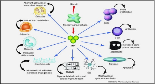
The common element: linkage of the four pathways
The link between activation of PKC, formation of AGEs, increased flux through the polyol pathway, a decrease in GSH, and formation of reactive oxygen species has been described by Nishikawa et al. (Nishikawa et al. 2000). This group used cultured endothelial cells to demonstrate that the hyperglycemia-induced increase in ROS production is caused through the TCA cycle (Nishikawa et al. 2000). Further, they reported that inhibition of mitochondrial respiration complex II (using the inhibitor rotenone) and oxidative phosphorylation (using the inhibitor CCCP and uncoupling protein-1 (UCP-1)), completely prevented the negative effects of hyperglycemia.
Real time quantitative PCR (RT-qPCR) validation of microarray results.
There are inherent limitations of microarray technology including non-specific hybridisation, poor hybridisation, false positive signals, background levels and issues raised by statistical analysis methods used for this technology. Consequently, independent confirmation of microarray results is frequently required for the publication of microarray results (Chuaqui et al. 2002; Firestein and Pisetsky 2002). As described by Chauaqui et al., (Chuaqui et al. 2002) there are a number of techniques that could potentially be used for validation, each with their own technical issues; however, the one most commonly used is RT-qPCR (Firestein and Pisetsky 2002). In this study and in all subsequent microarray studies RT-qPCR has been used as the microarray validation method.
Urine
Collection of 24hr urine enabled the measurement of TETA, metabolite and metal levels in urine in order to try and partly elucidate the metabolism of the drug in rodents. The method used to detect TETA and its metabolites (Section 2.7) has been validated by the School of Biological Sciences Reference lab for serum only and had not been tested in rodent urine before. There were initially some problems resolving the metabolites in the urine as MAT is at very high concentrations compared to DAT which is at very low concentrations. Further validation was performed by Reference lab technician Asma Othman that led to minor modifications being incorporated in order to ensure that all three compounds were detectable (Othman et al. 2007).
TETA and metabolite levels in urine as a function of time
The next step in the analysis was to identify what levels of TETA, MAT and DAT were being excreted in the urine and whether there were any differences comparable to those observed in the serum. Due to the collection of urine at the 10 Week (two weeks post-drug administration) and 15 Week (seven weeks post drug administration) time points, changes in excretion over time were incorporated into the analysis. In this analysis the three main effects, Status, Dose and Time were assessed. Data for TETA and MAT were analysed using a split-plot in time ANOVA as described in Section 2.12.5.1. One variable not accounted for in this analysis is the level of drug in the faeces of the animals as the metabolic cage study was not designed as a balance study.
Percentage of unmetabolised TETA in Serum and Urine
The results presented so far indicate that overall the diabetic animals have a greater amount of all three compounds (TETA, MAT and DAT) present in their urine at the Week 10 time point and more TETA and MAT present in their serum and at the Week 15 time point. The next step in the analysis was to identify in what proportion of the total excreted drug (TETA + MAT + DAT) unchanged or metabolised TETA was present and if there was an observable difference between diabetic and sham animals. This analysis helps to determine how much of the TETA absorbed was metabolised and how much remained as unmetabolised TETA. The results presented here focus on unmetabolised TETA only. Results are presented as a percentage of total drug in the urine (unmetabolised + metabolised TETA) so changes in unmetabolised TETA will reflect changes in metabolised TETA.
Table of Contents :
- Abstract
- Acknowledgments
- Table of Contents
- List of Figures
- List of Tables
- Abbreviations
- Chapter 1 General Introduction
- 1.0 Diabetes
- 1.1 Diabetic cardiomyopathy
- 1.1.1 Definition
- 1.1.2 Diabetic heart function assessed in animal models
- 1.1.3 Dysregulation of energy metabolism in the heart
- 1.1.3.1 Energy metabolism in the normal heart
- 1.1.3.2 Energy metabolism in the diabetic heart
- 1.2 Molecular mechanisms underlying diabetic complications
- 1.2.1 Pathway 1: An increase in Polyol-pathway flux resulting in a reduction in Glutathione
- 1.2.2 Pathway 2: Increased intracellular formation of advanced glycation end-products
- 1.2.3 Pathway 3: Activation of Protein Kinase C
- 1.2.4 Pathway 4: Increased flux through the Hexosamine pathway
- 1.2.5 The common element
- 1.3 Treatment of diabetic cardiomyopathy
- 1.3.1 Antioxidant therapies
- 1.3.2 Triethylenetetramine (TETA) as a treatment for diabetic cardiomyopathy
- 1.3.2.1 Current understanding of TETA pharmacology
- 1.4 Copper homeostasis
- 1.4.1 Normal copper homeostasis
- 1.4.1.1 Copper uptake into the cell
- 1.4.1.2 Copper distribution within cells
- 1.4.2 Copper and oxidative stress
- 1.4.3 Defective copper metabolism in diabetes
- 1.4.4 Copper and the development of cardiomyopathy
- 1.4.1 Normal copper homeostasis
- 1.5 Thesis Objectives
- 1.6 Experimental Approach
- 1.6.1 Streptozotocin-induced diabetic animal model
- 1.7 Summary
- Chapter 2 Materials and Methods
- 2.1. Animal model
- 2.1.1 Administration of Triethylenetetramine (TETA)
- 2.1.2 Metabolic cage 24hr urine collection
- 2.2 Isolation of RNA from the left ventricle of a rat heart
- 2.2.1 Tissue collection
- 2.2.1.1 Surgical procedure (Studies One and Two)
- 2.2.1.2 Perfusion and removal of left ventricle
- 2.2.2 RNA Isolation
- 2.2.2.1 RNA isolation from tissue using Qiagen MIDI Kit protocol
- 2.2.2.2 RNA isolation from tissue using a combined TRIZOL/Qiagen MINI method (Study Two)
- 2.2.1 Tissue collection
- 2.1. Animal model
- 2.3 Microarray Analysis
- 2.3.1 RNA
- 2.3.2 Ramaciotti Rat 10K Combo Slides
- 2.3.3 Amersham Codelink
- 2.3.4 Agilent
- 2.3.4.1 cRNA synthesis from total RNA
- 2.3.4.2 Hybridisation
- 2.3.4.3 Washing
- 2.3.4.4 Scanning and analysis of slides
- 2.3.5 Affymetrix
- 2.4 Real-time quantitative PCR validation
- Chapter 3 Pilot study to assess suitability of three commercially available microarray platforms for assessment of changes in gene expression
- 3.1 Introduction
- 3.1.1 Hybridising the probe to the array
- 3.1.2 Washing and staining of slides
- 3.1.3 Spot finding
- 3.1.4 Single versus competitive hybridisation
- 3.1.5 Cost and ease of use
- 3.2 Results
- 3.2.1 Spotted arrays from Clive & Vera Ramaciotti Centre for Gene Function Analysis
- 3.2.2 Amersham, Agilent and Affymetrix commercially available microarray systems
- 3.2.3 Definition of significant change in gene expression
- 3.2.4 Inter-platform variability
- 3.3 Discussion
- 3.3.1 Overview of the three commercially available systems
- 3.3.2 Correlation between Affymetrix and Agilent Systems
- 3.4 Conclusions
- 3.1 Introduction
- Chapter 4 Assessment of differences in the transcriptome between STZdiabetic and sham LV heart tissue at sixteen weeks
- Chapter 5 Characterization the physiological effects on and metabolism of TETA-disuccinate in diabetic and sham rats after eight weeks of treatment
- Chapter 6 Molecular changes in the left ventricle of diabetic and sham animals after eight weeks treatment with a high dose of TETA-disuccinate
- Chapter 7 Final Discussion and Conclusions
GET THE COMPLETE PROJECT
Investigation of diabetic cardiomyopathy and its treatment by copper chelation