Get Complete Project Material File(s) Now! »
Chapter 2. In vivo effects of epoxy omega-3 fatty acid analogues in a syngeneic glioma mouse model
Introduction
considerable amount of interest surrounding the health-promoting effects of omega-3 fatty acids has also led to their potential use as therapeutic agents against cancer (Fini et al., 2010; Miller & Gaudette, 1996; West et al., 2010). In vitro studies have demonstrated that these essential fatty acids can reduce the proliferation of various cancer cell lines (Begin, Ells, Das, & Horrobin, 1986; Liu et al., 2001; Yao et al., 2014), and also sensitise them to growth regulators and cytotoxic agents (Harvey et al., 2015; Hofmanová, Vaculová, & Kozubík, 2005). These anti-tumour effects have also translated to animal studies, where the treatment with omega-3 fatty acids was shown to reduce the growth of breast, colon, and endometrial tumours in vivo (Chen et al., 2014; Fluckiger et al., 2016; Pan et al., 2015). Fluckliger and colleagues (2016) documented that within the first five days of dietary DHA consumption, a significant reduction in the growth of s.c HCT-116 tumour xenografts in mice was seen compared to the control diet. Another study conducted by Pan et al (2015), revealed that a significant reduction of RL95-2 endometrial tumour xenografts in mice was positively associated with the levels of omega-3 fatty acid in the tissue when fed on a diet mixed with fish oil containing omega-3 fatty acids. However, these in vitro and in vivo results have not conclusively uncovered how omega-3 fatty acids achieved their anti-tumour effects. It is believed that their beneficial effects are mediated, in part, by their bioactive lipid mediators derived from, but not limited to, the oxygenation of omega-3 fatty acids. Numerous metabolic products can be formed to elicit a diverse range of biological processes including inflammation, vascular tone/function and angiogenesis (Node et al., 1999; Ulu et al., 2014; Yanai et al., 2014). The specific function of each metabolic product is still uncertain, however the biotransformation enzyme pathway, and the fatty acid which they are derived from, seem to have some significance. There are three different biotransformation enzymatic pathways for polyunsaturated fatty acids, and include the COX, LOX and CYP epoxygenase pathways. These pathways can give rise to products that have opposing effects depending on whether they are derived from omega-6 or omega-3 fatty acids (Funahashi et al., 2008; Hawcroft et al., 2012; Wada et al., 2007). For example, the metabolism of the omega-6 fatty acid, AA, via the COX pathway can result in the production of prostaglandin E2, which has been demonstrated to promote tumour growth, suppress immunity and stimulate tumour-promoting inflammation (Baryawno et al., 2008; Hoang, Allison, Murray, & Petrovic, 2015; Zelenay et al., 2015; Zheng, Ritzenthaler, Sun, Roman, & Han, 2009). In contrast, EPA is transformed to prostaglandin E3 under the same pathway, and acts as a partial antagonist, competing for the same receptor as prostaglandin E2 (Wada et al., 2007). Partially inhibiting prostaglandin E2’s tumour promoting effects, prostaglandin E3 has been shown to inhibit the proliferation of A549 lung cancer cells (Yang et al., 2004), and induce apoptosis of colorectal carcinoma cells and leukaemia stem cells (Hawcroft et al., 2010; Hegde et al., 2011).
Of the three enzymatic biotransformation pathways, the least studied to date is the CYP enzyme pathway. This pathway produces epoxy fatty acid metabolites (VanRollins, Frade, & Carretero, 1989) and again, depending on their parent fatty acid, they have been associated with a wide range of effects including in vitro cancer cell aggressiveness, high blood pressure, vascular reactivity (Capdevila, Falck, & Harris, 2000; Lopez-Vicario et al., 2015; Wei et al., 2014). Recently, Zhang and colleagues (2013) chemically synthesised five different regioisomers of EDP, including 19,20-EDP, which consists of an epoxy group at the omega-3 position. They examined 19,20-EDP in a in vivo syngeneic Met-1 breast cancer model and demonstrated that when co-administrated with the sEH inhibitor, tAUCB, significant reductions in tumour volume and tumour weight were observed. Without sEH inhibition, 19,20-EDP failed to elicit significant anti-tumour effects (Zhang et al., 2013). This is because epoxy omega-3 fatty acids can be further metabolised to their corresponding, less active, diols by the sEH (Fleming, 2008). Epoxides may have therapeutic benefits in various inflammatory conditions and cancer, hence their stabilisation by pharmacologically inhibiting sEH has been of academic and commercial interest with some inhibitors currently being tested in clinical trials (Imig et al., 2005; Lazaar et al., 2015; Watanabe & Hammock, 2001). Unfortunately, the inhibition of sEH will also stabilise epoxy omega-6 fatty acids, which has been shown to increase their effects to promote tumour growth and metastases in animal tumour models (Panigrahy et al., 2012). Together these findings suggested that the epoxy metabolites derived from omega-3, but not omega-6 fatty acids, may have beneficial effects against cancers. The presence of olefinic double bond at the omega-3 position of omega-3 fatty acids, in contrast to the absence in their omega-6 counterparts, has been proposed to be of biological significance for the anti-tumour activity of these fatty acids (Cui et al., 2011).
As bioactive lipid mediators are generated from free fatty acids released from phospholipids (Hammond & O’Donnell, 2012), dietary behaviours that influence phospholipid composition can subsequently shape the assortment of lipid mediators produced (Abbott, Else, Atkins, & Hulbert, 2012; Schuchardt et al., 2014). In the Western diet, omega-6 fatty acid predominates over omega-3 fatty acid intake, where the omega-6 to omega-3 fatty acid ratio is approximately 16:1 (Chajes & Bougnoux, 2003; Simopoulos, 2002a). As a result, the relative abundance of omega-6 fatty acid favours omega-6 derived oxylipins being formed. Suggestions of a shift towards lower intake of omega-6 fatty acids and higher intake of omega-3 fatty acids have been discussed to promote human health (Murray, 2013; West et al., 2010). An alternative strategy is to design and develop analogues of mediators derived from omega-3 fatty acids that have shown favourable effects against cancer. Recently, the synthesis and investigation of a series of analogues based on the omega-3 fatty acid-derived metabolite, prostaglandin J3, has identified a lead compound that possesses high potency against numerous cancer cell lines, including the U251 glioblastoma cell line (Nicolaou et al., 2016). Thus far, these analogues have shown to selectively eradicate leukaemia stem cells in vivo, in a potent pro-apoptotic fashion and may represent a new class of chemotherapeutic agents (Hegde et al., 2011).
The development of a novel class of anti-tumour agents based on epoxy omega-3 fatty acid has also been initiated, where substituting the omega-3 epoxide with a urea group has been utilised to improve stability while retaining pharmacological effect (Dyari et al., 2014; Falck et al., 2009; Falck et al., 2011). The urea bioisosteres have been shown to maintain anti-proliferative effects against the human breast cancer cell line, MD-MBA-231 (Dyari et al., 2014) and have provided the basis for further structural modification to enhance the potency of these agents. A number of urea bioisosteres of epoxy omega-3 fatty acids, similar to the one described by Dyari et al (2014) have also been examined in vivo. CUT-EE (Figure 2.1), is an analogue that was shown to exert anti-tumour activity against MD-MBA-231 breast tumours in immune-deficient
Balb/c nu/nu mice at a dose of 10 mg/kg i.p (Prof M. Murray, personal communication). Following their promising findings, experiments with the another analogue, C29 (Figure 2.1), was also found to decrease the size of primary tumours reduce the number of metastases in the MD-MBA-231 xenograft model at a daily dose of 40 mg/kg i.p (Rawling et al., manuscript submitted for publication, Prof M. Murray, personal communication). These effects were also observed in experiments using a daily dose of C29 at 20 mg/kg, but to a lesser extent when compared to the higher 40 mg/kg dose (Prof M. Murray, personal communication). Based on these encouraging findings, the work in this chapter set out to evaluate the activity of these novel agents, C29 and CUT-EE, against the mouse GL261 glioma model.
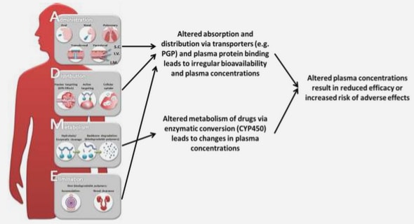
Currently, there is a need for more effective therapies for patients with glioma, which is the most common and aggressive type of all adult brain cancers (Dolecek et al., 2012). The multi-modal approach, which involves surgery, radio- and chemo-therapy, offers patients with glioblastoma a median survival of 14.6 months and a 5-year overall survival rate of 9.8% (Stupp et al., 2009; Stupp et al., 2005). Despite treatment, tumours inevitably recur within close proximity to the initial tumour. Scientists designing and developing effective agents to treat brain tumours encounter the challenge of delivering the agents across the BBB. Fatty acids, particularly AA and DHA, are found in brain tissue, and because this organ cannot synthesise these essential fatty acids necessary for its normal function (Demar et al., 2005; McCann & Ames, 2005), they are obtained via the systemic circulation through the diet. Brain uptake of fatty acids has been well established through numerous studies (Freund Levi et al., 2014; Ouellet et al., 2009; Pardridge & Mietus, 1980; Washizaki et al., 1994). It was anticipated that the mechanisms of natural fatty acid brain uptake could be exploited with the intention to overcome the BBB to treat brain tumours.
The GL261 tumour was originally a carcinogen-induced (methylcholanthrene) murine glioma model developed in immunocompetent C57BL/6 mice (Seligman, Shear, & Alexander, 1939). Later, the tumours were subsequently dissected, grown in culture for further use in transplantation studies with syngeneic mice. This method of transplantation enables researchers to more accurately and reproducibly grow tumours in their location of origin. Owing to its invasive, angiogenic and morphological characteristics that resemble human glioblastoma multiforme, the GL261 glioma is widely used as a model in brain cancer research (Hanihara et al., 2015; Newcomb et al., 2006; Shapiro, Ausman, & Rall, 1970; Szatmari et al., 2006; Wainwright et al., 2014; Yung et al., 2014; Zagzag et al., 2000). In this chapter, the anti-tumour effects of the novel epoxy omega-3 fatty acid analogues, C29 and CUT-EE, were investigated using the GL261 cell line in an ectopic s.c tumour mouse model. S.c tumour models are extensively used in the preclinical setting of cancer research to examine the effectiveness of investigational agents (Ching et al., 2002; Henare et al., 2012; Yung et al., 2014). The s.c “flank” tumours are located in a convenient and exposed site to where researchers can access and measure tumour size easily, however, the tumour grown at this location are not protected by the and therefore limits its usefulness of this model when investigating new treatments for tumours residing in the brain. A more favourable model that would more closely recapitulate the clinical setting would be to use mice with stereotactically implanted tumours in the brain when examining and developing specific agents against brain tumours. The i.c tumour model has been previously established in our laboratory and is used frequently as a model for brain tumour research (Hanihara et al., 2015; Newcomb et al., 2006; Yung et al., 2014), and although the i.c implantation of tumour cells involves an invasive and disruptive procedure where BBB integrity could be lost, multiple publications have concluded that the BBB is still functional at early time points after implantation. Considering this, the anti-tumour effects of the novel epoxy omega-3 fatty acid analogues were also examined against an orthotopic i.c tumour mouse model. In many cases, the GL261 luciferase transfected cell line has been utilised to allow for non-invasive monitoring of tumour establishment, growth and response to treatment in live animals (Goldhoff et al., 2008; Kindy, Yu, Zhu, Smith, & Gattoni-Celli, 2016). As brain tumours cannot be non-invasively visualised or confirmed without expensive and time consuming techniques such as magnetic resonance imaging, this experimental chapter used a GL261 cell line that has been transfected with the luciferase gene, named GL261-luc2.
Aims
The aims were:
To examine the in vitro activity of epoxy omega-3 fatty acid analogues against the GL261-luc2 cell line.
To determine the anti-tumour activity of epoxy omega-3 fatty acid analogues in the ectopic s.c GL261-luc2 mouse glioma model.
To determine the anti-tumour activity of epoxy omega-3 fatty acid analogues in the orthotopic i.c GL261-luc2 mouse glioma model.
Materials and methods
Omega-3 epoxy fatty acid analogues with toluene substituents at the C-2 position, and ethyl esters at the carboxylic acid end were synthesised at the School of Pharmacy, Graduate School of Health, The University of Technology Sydney, Australia. The structural identities were determined by mass spectroscopy, 1H and 13C nuclear magnetic resonance, and the purity was confirmed by elemental analysis. Unless otherwise stated, all other chemicals were commercially available and of analytical grade. Water used in all experiments was purified by filtering through ion exchange columns and a 0.22 μm filter (Milli-Q Purification System, Millipore Corporation, Bedford, USA).
Animals
All animal experiments were done following protocols approved by the University of Auckland Animal Ethics Committee, and met standards required by the United Kingdom Co-ordinating Committee on Cancer Research guidelines. C57BL/6, and B6 Albino female mice (1823 g) were used and housed under constant temperature (20°C), humidity, and lighting (12 h light per day). The B6 Albino strain is a spontaneous mutant of the C57BL/6 strain. The homozygous mutation in the tyrosinase gene results in a white (albino) coat colour rather than black.
GL261-luc2 glioma cell line
The GL261 mouse glioma cell line was obtained from the National Institute of Health and Sciences, USA. To enable the glioma cells to be detectable using an optical image for i.c tumour detection, the GL261 cell line stably-expressing a luciferase gene (F279-V5 luciferase 2, Invitrogen, Carlsbad, CA, USA) (Yung et al., 2014) was used and maintained in minimal essential medium (MEM) alpha media (GIBCO, Grand Island, NY, USA) supplemented with 10% heat-inactivated fetal calf serum under puromycin (1μM) selection at 37°C in a humidified atmosphere containing 5% CO2. These cells were subsequently called GL261-luc2. Cells were passaged using 0.05% trypsin/EDTA and suspended in antibiotic- and serum-free medium before implantation in animals. After revival from liquid nitrogen storage, cells were cultured for no more than 8 passages before implantation in animals.
In vitro cell viability assay
GL261-luc2 cells were cultured and passaged similarly to what was previously described in section 2.3.2. To optimise the number of cells required for cell viability assays, cells were titrated in 96 well plates and allowed to grow over 24 h duration. After the allocated time, cell viability was determined with the addition of 20 μl of 3-(4,5-Dimethylthiazolyl-2)-2,5-diphenyltetrazolium bromide (MTT) at 5 mg/ml in phosphate buffered saline (PBS) (filter sterilised). MTT is reduced by cellular metabolic enzymes and, in defined conditions, reflect the number of cells (Mosmann 1983). The cells were incubated with MTT for 30 min at 37°C, and the plates were subsequently centrifuged at 300 x g for 5 min before the supernatant was removed. DMSO (100 μl) was added to each well and shaken on plate shaker for 10 min to solubilise formazan crystals and optical density of wells were determined at 550 and 650 nm using a microplate reader (SpectraMax M2, Molecular Devices, Sunnyvale, CA, USA). To determine the percentage of the DMSO that could be used without influencing cell viability for drug-response experiments, various percentages of DMSO (v/v) in cell growth medium were incubated with GL261LUC2 cells and cell viability was determined similarly to above. For drug treatment experiments, CUE, CUT, C29, iPUE, iPUT, and CUT-EE were dissolved in DMSO to give stock solutions of 10 mg/ml. These were further dissolved in medium to give a concentrations that was twice the required final concentration in wells. Vehicle controls using DMSO without analogues were also prepared similarly. After cells were pre-incubated in wells for 30 min a volume of 100 μl drug solution was added to 100 μl cell solution in wells. After a 24 h incubation with the agents, cell viability was determined with the methods described above. The response to agents were calculated using absorbance measured at 550 nm minus background absorbance at 650 nm. This value was then calculated as a percentage of vehicle control, where control values was assigned with a cell viability of 100%. Curves were fitted using nonlinear regression and concentrations required to inhibit 50% of the control cell viability (IC50) were determined by interpolation from y-axis values.
Tumour implantation
For s.c tumours, 1 x 10⁵ GL261-luc2 cells suspended in unsupplemented medium in a volume of 100 µl were injected s.c into the left flank of C57BL/6 mice using a 0.5 ml, 29-gauge insulin syringe (BD, San Jose, CA, USA). For i.c tumour implantations, B6 Albino mice were anaesthetised with ketamine (100 mg/kg) and xylazine (10 mg/kg), and animal fur at the surgical site was removed by hair removal cream. Mice were placed in a stereotaxic frame (Kopf Instruments, Tujunga, CA, USA) and a small incision on the scalp was made down the anterior posterior axis to expose the bregma. A small bur hole was made at the coordinate’s −0.1 mm anteroposterior, +2.3 mm mediolateral from bregma. A Hamilton syringe containing GL261-luc2 at a concentration of 5 x 10⁷ cell/ml was placed in the bur hole and lowered slowly to a depth of -3.2 mm. A total volume of 2 μl (1 x 10⁵ cells) cell suspension was delivered into the caudate/putamen of the mouse brain at a rate of 0.5 μl/min using a micro-pump (World Precision Instruments, Sarasota, FL, USA)-controlled Hamilton syringe. After cell delivery, the syringe was kept in place for 2 min, and subsequently raised 1.5 mm and left in place for a further 1 min. Upon withdrawal the incision was sutured and temgesic (100 μg/kg) was administered s.c along with fluid replacement therapy (10 ml/kg, 0.9% sterile saline). Animals were monitored until they were fully recovered from anaesthesia.
Bioluminescence imaging
In vitro bioluminescence (BLI) from GL261-luc2 was quantified using a black 96-well plate containing control wells; media, cells, and D-luciferin only (Gold Biotechnology, Inc, Olivette, MO, USA), along with a titration of GL261-luc2 cells (5×10³, 1×10⁴, 5×10⁴, 1×10⁵, 5×10⁵ cells/well) containing 15 mg/ml D-luciferin in PBS. The plate was placed inside the IVIS Kinetic optical imager (PerkinElmer Waltham, MA, USA) immediately after adding the substrate and a BLI image was acquired. B6 Albino mice were used for the i.c tumour model as they lack dark dermal pigmentation, which may sequester BLI signal intensity. In treatment studies, the presence of i.c tumours was determined using BLI imaging before initiating treatment. Non-tumour bearing mice and mice implanted with i.c GL261-luc2 were anaesthetised with isoflurane gas (Lunan Pharmaceutical Group Co., Shandong, China). On imaging day, D-luciferin was made fresh in PBS at 30 mg/ml and administered i.p at 150 mg/kg. Animals were then placed in the IVIS imaging system and the BLI was measured using the system parameters: exposure time = auto, binning = medium, F/stop = 2, EM gain = 100. A series of images, 1 min apart, were captured between 5 and 18 min after substrate injection. This time period was appropriate to capture maximum BLI from tumours, which was previously determined to be approximately 10-12 min after substrate administration (Yung et al., 2014). Analysis of images was performed using the Living Image®3.2 software (PerkinElmer Waltham, MA, USA) by identifying a region of interest over the brain tumour region and obtaining the BLI as photons/s. The XFOV-24 lens was installed to image multiple animals (up to 5) (Caliper Life Sciences, Hopkinton, MA, USA). The maximum BLI signal value during the time period was corrected for the background/baseline signal level and used to monitor tumour growth over time. Mice that were not implanted with cancer cells were used as a BLI background/baseline control.
Table of contents
Abstract
Acknowledgements
Table of contents
List of figures
List of tables
List of abbreviations
Chapter 1. General introduction
1.1 Brain cancer
1.2 Treatments for glioma
1.3 The blood-brain barrier
1.4 Overcoming the blood-brain barrier for drug delivery
1.5 Brain uptake of fatty acids
1.6 Fatty acids and cancer
1.7 Summary and aims of thesis
Chapter 2. In vivo effects of epoxy omega-3 fatty acid analogues in a syngeneic glioma mouse model
2.1 Introduction
2.2 Aims
2.3 Materials and methods
2.4 Results
2.5 Discussion
Chapter 3. Development and validation of a bioanalytical method for epoxy omega-3 fatty acid analogues in mouse tissues
3.1 Introduction
3.2 Aims:
3.3 Materials and methods
3.4 Results
3.5 Discussion .
Chapter 4. Tissue distribution and pharmacokinetics of epoxy omega-3 fatty acid analogues in mice
4.1 Introduction
4.2 Aims
4.3 Materials and methods
4.4 Results
4.5 Discussion
Chapter 5. Concluding discussion
5.1 Future direction
5.2 Conclusion
Chapter 6. List of references
GET THE COMPLETE PROJECT
Preclinical studies of epoxy omega-3 fatty acid analogues for treating glioma