Get Complete Project Material File(s) Now! »
Physics of diffusion magnetic resonance imaging (dMRI)
Magnetic resonance imaging (MRI) is a non-invasive technique based on the principle that, in presence of a constant external magnetic field B0, a set of particles that have non-zero spin, will see their magnetization align along the direction parallel to B0. For example, if we consider water protons, they have spin 1{2. Under a constant magnetic field B0, these nuclei have two state of energies, B0 and B0, with being the nuclear magnetic moment. The energy difference corresponds to a resonant or Larmor frequency w B0; (1.4) where is called gyro-magnetic ratio, and for water protons is equal to 2:675 108rad T 1s 1 (see [63]). At thermal equilibrium, the difference of state populations creates a net magnetization which is oriented along the direction of the magnetic field and is “precessing” around this direction at frequency w. Nevertheless, this net magnetization has very low intensity compared to the intensity of B0. In order to be able to measure it, the idea is to “flip” this magnetization on a transverse plane (or simply on a plane which forms an angle with the direction of the applied magnetic field, where the amplitude depends on the application). To do so, a periodic magnetic field B1, rotating in the transverse plane at the Larmor frequency w, is applied for short time. This magnetic field B1 is called “RF pulse” because it consists in an electromagnetic radiation with a precise radiofrequency applied for a short time. Once B1 is turned off, the spins tend to come back to their initial condition via a phenomena called “relaxation” and we are then able to measure a signal which represents the loss in net magnetization. Since the magnetization can be represented as a vector, we decompose it into two components, parallel and perpendicular to the direction parallel to B0, and we distinguish between two relaxation phenomena characterized by two times: T1 which gives information on how fast the longitudinal magnetization comes back to the initial value and T2 which gives information on how fast the transversal magnetization goes to 0. In general, signals would suffer additional suppression due to dephasing from external field inhomogeneities. A rephasing can be achieved by an additional RF pulse application, where the basic idea is to flip the phase of the spins by 180 in the transverse plane (for more details see for example [28, 90]). Following the 180 flip the dephasing is reversed, and the phases refocus at what is called echo time TE. The signal is then acquired at time TE. To be able to localize the signal and create an image, also gradient fields are applied. A gradient field is a magnetic field which varies in space. In particular, three linear gradients are used: the “slice-selection” gradient (Gsl) to select a region in which we want to excitate the spins, the “frequency” and “phase-encoding” gradients (Gx and Gy) to generate the signal in the Fourier domain. These three gradients are also called “imaging gradients”. The slice-selection gradient Gsl is applied at the same time as the RF pulse and thanks to it we select a volume of spins that precess at the Larmor frequency determined by B0, while the surrounding spins are precessing with different frequency. Lets suppose for simplicity that B0 is directed along the z-direction and that B1 flips the spins in the transversal plane xy. After spins are excited (i.e. after the application of Gsl), new linear gradients are applied along the two in-plane directions (x and y) defining the excited slice. The resulting linear changes in magnetic field create corresponding linear changes in precessional frequency (i.e. the frequency w is determined by the sum of B0 and the linear gradients). As these frequencies vary over time with gradient duration, the excited spins accumulate also phases based on their location px; yq and the integral in time of the applied gradients (Gx and Gy).
Some biological applications and advanced acquisition
We have seen that the dMRI signal is the transverse magnetization in a volume of tissue called a voxel. The application of diffusion-encoding gradients causes an attenuation of the magnetization due to the dephasing of spins (water protons) from the incoherent motion of water. Since in biological tissues water diffusion is not free, and is instead strongly affected by the local environment that can contain hinderance to water displacement such as cell membranes and macromolecules, dMRI can reveal information about the tissue microstructure even though the signal is collected on a macroscopic level (voxel). In biological tissues, the image contrast in water proton diffusion magnetic resonance imaging is given by the difference in the average water displacement due to the difference in diffusion between imaged tissues at different spatial positions [106]. Since the first diffusion MRI images of normal and diseased brain in [107], an early major application has been the study of acute cerebral ischemia in stroke [131, 187]. In the brain, dMRI has been also used to detect a wide range of physiological and pathological conditions, including tumors [125, 166, 174, 181], myelination abnormalities [80, 108], connectivity [105], as well as in functional imaging [110, 111].
There are multiple ways to display contrast using dMRI. Although this is not the focus of the thesis, for completeness, and in order to give the reader some basic references on the applications of this technique, here I report a short list of some of the most popular and used ways to display contrast using dMRI. An early contrast is the simplest one, where the intensity of each pixel of the image represents the magnitude of the transverse magnetization for a certain choice of diffusion-encoding gradient strength and direction. This contrast is the basis of diffusion-weighted imaging (DWI), which was shown to be more sensitive to early cellular changes after a stroke than more traditional MRI measurements (see for example [81, 82, 96, 131, 188] and Figure 1.3). DWI is most applicable when the tissue of interest results in isotropic water diffusion displacement, for example in the brain grey matter.
The apparent diffusion coefficient (ADC)
An important quantity measured in dMRI is the apparent diffusion coefficient (ADC) or effective diffusion coefficient (Deff), with the former term often used by medical researchers and some MR physicists. The latter is the preferred term among mathematicians and some physicists. In this thesis, we will use both ADC and Deff, but we distinguish them in the following way. (The choice we made, described below, is somewhat arbitrary and we hope that it does not detract from the clarity of the thesis.)
In this thesis, the ADC will be used to designate the quantity that includes the contributions from all the geometrical compartments in a volume of interest, whereas Dneff will be used to designate the quantity that accounts for the contribution from a particular compartment n, within the volume of interest. (For example, all the spherical cells could comprise compartment 1, all the cylinder cells could comprise compartment 2, and the extra-cellular space compartment 3. The union of these three compartments make up the total volume.) As a consequence, if we are considering just one compartment, then ADC Deff, whereas if we are considering N ¡ 1 compartments, and exchange between the compartments is negligible, the ADC is.
Table of contents :
Introduction (en français)
Introduction
1 Diffusion magnetic resonance imaging
1.1 Diffusion
1.2 Physics of diffusion magnetic resonance imaging (dMRI)
1.3 Some biological applications and advanced acquisition
2 Mathematical models
2.1 The microscopic Bloch-Torrey model
2.1.1 Probabilistic interpretation
2.1.2 Important length scales
2.1.3 Solutions of Bloch-Torrey equation
2.1.4 Narrow pulse approximation
2.1.5 The apparent diffusion coefficient (ADC)
2.1.6 Gaussian phase approximation
2.2 Approximate models
2.2.1 Short-time approximation
2.2.2 Long-time approximation
2.2.3 Multi-compartments models
2.2.4 Geometric models
3 Finite pulse Kärger and Kärger Models
3.1 Description of the models
3.1.1 Finite Pulse Kärger model
3.1.2 Kärger model
3.2 Convergence of the Kärger model to the FPK model
3.2.1 Asymptotic expansion in
3.2.2 Error estimates
3.3 Kurtosis formula for FPK model
3.4 Numerical results
3.4.1 Trapezoidal PGSE
3.5 Conclusions
4 New homogenized model of time-dependent ADC (H-ADC)
4.1 Problem setting
4.1.1 Bloch-Torrey equation
4.1.2 Periodicity length
4.2 An asymptotic model
4.2.1 Transformed Bloch-Torrey equation
4.2.2 Choice of scaling
4.2.3 Asymptotic model corresponding to
4.2.4 Asymptotic dMRI signal model and its ADC
4.3 Numerical results
4.3.1 Convergence
4.3.2 Time-dependent ADC
4.4 Comparison between the new asymptotic model and the linearized model
4.4.1 Convergence
4.5 Conclusions
5 Other scalings for small b-values
5.1 General equations
5.2 Parameters limitations
5.3 Diffusion time scale comparable with the cell size 1
5.4 Diffusion time scale shorter than the cell size 2
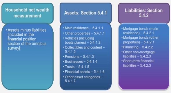
5.4.1 Moderate intensity:
5.4.2 Relatively strong intensity: 1
5.4.3 Very strong intensity: 3
5.5 Mini space dependent for 2
6 Time-dependent ADC inside cells
6.1 Effective diffusion coefficient in finite domains
6.2 Solution of the model
6.2.1 Eigenfunctions representation (finite pulse long-time formula, FPLT)
6.2.2 Layer potential representation (short pulse short-time formula, SPST)
6.2.3 Mixed approximation (short pulse long-time formula, SPLT)
6.3 Averaging Deff over multiple diffusion directions
6.4 Numerical results
6.5 Conclusions
7 On the inverse problem
7.1 Analysis based on the new formulas for ADC
7.1.1 Finding the surface over volume ratio in the short-time limit using SPST .
7.1.2 Finding the surface over volume ratio and the eigenvalues
7.2 On the determination of radii distributions
7.3 On the detection of the fibers orientations
Conclusions and Perspectives
A Physical background
A.1 Spatial and Contrast resolution
A.1.1 Nuclear Magnetic Resonance
A.1.2 Spin angular momentum
A.1.3 Energy Levels
A.2 Generating the signal
A.2.1 Resonance
A.2.2 Relaxation
A.2.3 Contrast
A.3 Gradient magnetic field
A.3.1 Slice selection
A.3.2 Frequency and Phase encoding
A.4 Diffusion MRI
Acknowledgements / Remerciements
Bibliography