Get Complete Project Material File(s) Now! »
Theoretical background
This chapter describes the theories and previous research found in the literature that covers the subjects of this thesis. The trabecular structure and its pore topology and how the structure affects the mechanical properties is first presented followed by information about Selective Laser Melting, the material used; Ti-6Al-4V and last previous research.
Trabecular structure
Trabecular bone is the inner part and the cortical bone is the outer part of the natural bone, they belong to porous materials with interconnected voids [3]. The trabecular bone is found in the vertebral bodies of the axial skeleton and at the end of the long bones of the appendicular skeleton. It has a porous structure pattern. This complex patterns assists to maximal strength for minimum mass for the skeleton. The trabecular network has both rod-like and plate-like structures with interconnected voids, which has a random structure and can be found in more or lesser proportion depending on the skeletal site. The trabecular bone provides, due to high mineral surface area, an extensive substrate on which cellular interaction with bone mineral material can occur [4].
Pore architecture
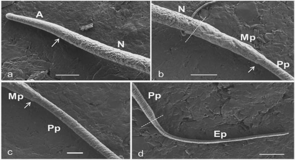
With additive manufacturing it is possible to obtain similar design, which is interconnected pores as natural bone i.e. pore size and pore distribution (Figure 2). Titanium is known for its good biocompatibility since decades and it is already used as material for medical implants. In form of a trabecular structure it can lead to the development of innovative orthopaedic prostheses [3]. The morphology of porous materials can be both stochastic (random) and ordered (regular) depending on the manufacturing settings [5]. The trabecular structure has an open-cell structure, meaning it is permitting flow of fluid inside the scaffold, the other type of porosity is closed-cell porous, that can be found inside the structure, i.e. it cannot permit flow of fluid [6]. There are criteria that porous biomaterials should be able to achieve for bone replacement. These criteria include: filling of bone defect voids, the pore architecture and the pore interconnectivity should promote osseointegration and sufficiently good mechanical properties to support physiological loading. The function of the porous biomaterial structure needs to consider the microarchitecture like pore shape and size,cell topology and porosity fraction, both of these have a high impact on the osseointegration, interface strength and mechanical behaviour. For optimal osseointegration the pore size should be between 50 μm and 800 μm and have a porosity higher than 50% [7]. With a porosity of 75% to 80% an increase in fixation strength can be obtained and it is needful to have interconnectivity between the pores in order to permit osseointegration [8]. There are limits of the pore size due to the biological factors, pores larger than 700 μm can promote osseointegration but at reduced rates and volumes of bone ingrowth. Pore size less than 100 μm loses the support of the growth of capillaries and also allow the bone cells to bridge and close pores .
1 Introduction
1.1 BACKGROUND
1.2 PURPOSE AND RESEARCH QUESTIONS
1.3 RESEARCH APPROACH
1.4 DELIMITATIONS
1.5 OUTLINE
2 Theoretical background
2.1 TRABECULAR STRUCTURE
2.1.1 Pore architecture
2.1.2 Biomedical division
2.2 SELECTIVE LASER MELTING
2.2.1 The process
2.2.2 Characteristics
2.2.3 Sustainability perspective
2.3 TI-6AL-4V
2.3.1 Composition and microstructure
2.3.2 Mechanical behaviour
2.4 PREVIOUS RESEARCH IN THE FIELD
3 Method and implementation
3.1 PRELIMINARY WORK
3.1.1 Porosity fraction
3.1.2 Morphological analysis
3.2 SPECIFICATION OF SPECIMENS
3.2.1 Batches
3.2.2 Structures
3.2.3 Porosity fraction, microstructural analysis, compression test and morphological analysis
3.2.4 Friction test
3.2.5 Tensile test
3.2.6 Abrasive test
3.3 METHOD PLANNING
3.4 PHASE Ⅰ
3.4.1 Porosity fraction
3.4.2 Microstructural analysis
3.4.3 Compression test
3.4.4 Morphological analysis
3.4.5 Evaluation phase Ⅰ
3.5 PHASE Ⅱ
3.5.1 Friction test
3.5.2 Tensile test
3.5.3 Abrasion test
4 Findings and analysis
4.1 PHASE Ⅰ
4.1.1 Porosity fraction
4.1.2 Microstructural analysis
4.1.3 Compression test
4.1.4 Morphological analysis
4.2 PHASE Ⅱ
4.2.1 Friction test
4.2.2 Tensile test
4.2.3 Abrasive test
5 Discussion and conclusions
5.1 DISCUSSION OF METHOD
5.1.1 Preliminary work
5.1.2 Porosity fraction
5.1.3 Compression test
5.1.4 Morphological analysis
5.1.5 Friction test
5.1.6 Tensile test
5.1.7 Abrasive test
5.2 DISCUSSION OF FINDINGS
5.2.1 Porosity fraction
5.2.2 Compression test
5.2.3 Morphological analysis
5.2.4 Friction test
5.2.5 Tensile test
5.2.6 Abrasive test
5.3 CONCLUSIONS
5.4 FUTURE WORK
6 References
7 Search terms
8 Appendices