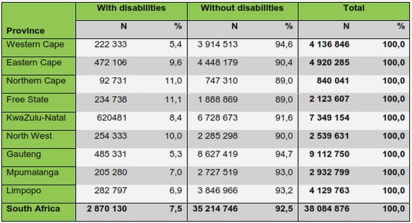
Get Complete Project Material File(s) Now! »
Synchronization in the heart
In order to achieve efficient pumping action, billions of cardiac cells have to work together in a synchronized fashion. Each heartbeat is initiated by a pulse of electrical excitation that begins in a group of specialized pacemaker cells and subsequently spreads throughout the heart. This electrical impulse is made possible by the electrochemical gradient that exists across the surface membrane of each myocyte. At rest, the membrane is selectively permeable to K+ ions, and the electrochemical potential inside the myocyte is negative with respect to the outside.
During electrical excitation, there is an influx of current from the neighbouring tissue, the membrane becomes permeable to Na+ and the electrochemical potential reverses or depolarizes. The changes in Na+ and K+ lead to osmolarity fluctuations which causes Ca2+ ions to reorganize across the cell wall and inside the cytoplasm. The movement of Ca2+ into the cell cytoplasm activates the contractile machinery, a process that, when it happens en masse, causes the heart chamber to contract and expel blood. The depolarization is limited by the restoration of the negative membrane potential, this allows the chamber to relax and fill with blood for the next cycle.
Because the heart beat is critically dependent on the proper movement of ions across the surface membrane, disorders of ion channels or channelopathies make up a key group of heart diseases. Channelopathies predispose individuals to disturbances of normal cardiac rhythm. When the heart beats too slowly (bradyarrhythmias) or so rapidly that it cannot fill adequately (tachyarrhythmias), this leads to circulatory collapse and, in the extreme case, death. Several different genetic and acquired channelopathies can cause such arrhythmias.
This chapter provides a detailed introduction to the excitation mechanisms and interaction of these mechanisms with other cellular processes like contraction and metabolism.
Genesis of the myocyte excitation
Normal cardiac excitability results from the balance of depolarizing and repolarizing ionic cur-rents. Each ionic current can be distinguished by its ionic selectivity and time course. These properties are conferred by specific transmembrane proteins called channels or gates. Figure 3.1A depicts the principal channels and transporters in myocytes. Depolarization is mediated by channels or gates that enable positively charged ions such as Na+ or Ca2+ to enter the cell (or negatively charged ions, usually Cl , to exit) and vice versa for repolarization.
Many proteins have evolved to mediate the selective flux of ions across biological membranes. Proteins called Ion channels depend on the electro-chemical gradient across the membrane. Ion channels are usually selective for one type of ion over all others. When these ion channels open, they bias the membrane potential of the cell towards the equilibrium potential of that ion. For example, K+ channels steer the cell towards 90 mV, while the opening of Na+ channels forces the potential towards positive levels (+40 mV ).
Thus, one can visualize each cell as a dipole that is either positive or negative depending on the relative balance of the cell’s complement of ion channels, and whether or not those channels are open at any given time. The resulting electrical signal is called the action potential. A typical action potential profile of a ventricular myocyte is shown in the centre of Figure 3.1B.
The action potential has a sharp depolarizing upstroke which gives way to a sustained, slowly decaying plateau and eventually repolarizes. Figure 3.1A shows the depolarizing ionic currents, which are generated by the channels (and a single gate, the Na+=Ca2+ exchanger), that underlie the electrical signal. Ion channels allow the passive movement of ions down their respective concentration gradients. When K+ channels open during repolarization, K+ exits from the cell, conversely, during depolarization Na+ and Ca2+ enter the cell.
The ionic gradients are maintained by active energy-consuming processes carried out by pro-teins such as the Na+=K+ATPase. The Na+ channels and Ca2+ channels favour depolarization. Each opens quickly in response to a voltage stimulus causing further depolarization, and then closes despite maintained depolarization– a process known as ‘inactivation’. Under normal condi-tions, Na+ channels inactivate quickly and completely and very rarely re-open during the course of the action potential. Calcium channels on the other hand inactivate less rapidly and less completely and feature prominently in maintaining the plateau phase. The gates involved in the repolarizing mechanism consist of various types of K+ channels. The K+ currents are shown in Figure 3.1C.
Voltage sensitive mechanisms
Ion channels and gates with activation/inactivation dynamics determined by the ionic concentra-tion and the potential difference across the membrane surface are primarily responsible for the observed electrophysiological behaviour. Figure 3.2 shows a schematic of the voltage sensitivion dynamics and excitation propagation in cardiac myocytes. The ionic fluxes generated during the action potential generate observable currents and are tightly regulated in a normal myocyte.
Ionic currents and ion channels
This section discusses some of the predominant ionic currents and the ion-channels that are responsible for these currents.
Sodium currents and its components.
The sodium current is primarily responsible for the upstroke and conduction of the action po-tential. The cardiac Na+ current is manifested by the sodium ion channel which is encoded by the SCN5A gene. The activation of the current is regulated by the membrane voltage; the inactivation of the current seems to be initiated by the activation process. Sodium channels have three types of states: deactivated (closed), activated (open), and inactivated (closed). Channels in the deactivated and inactivated state can be thought of being blocked on their intracellular side by a « door », which opens in response to stimulation and hence opens the channel.
The gate quickly closes after a large influx of Na+ ions. During an action potential the channel remains inactivated for a few milliseconds after depolarization. The inactivation is removed when the membrane potential of the cell repolarizes following the falling phase of the action potential. Voltage based activation of the Na+ current can be viewed as a manifestation of all the Na+ ion channels in the cell. The Na+ channels go through the following state transition sequence.
Voltage sensitive mechanisms
Activated, Deactivated, Inactivated. Thus, the strength of the response is proportional to the available inactivated channels. The repolarization membrane voltage and the duration that the cell remains at that voltage determine the number of channels in the inactivated state. This causes the change in depolarization potential with activation frequency.
Mutations and alterations (by drugs/toxins) to the inactivation gate lead to changes in the action potential duration and shape. Slowing of the inactivation process, observed in LQT3 syndrome, leads to long action potentials [11]. Reduction in Na+ current due to changes in the inactivation process has been shown to lead to Brugada syndrome [12]. In some instances conduction block due to low Na+ current has also been observed [13].
Potassium currents and its components
Unlike the sodium ion, which has only one associated current, there are many potassium ion related currents. The primary role played by these currents is to aid in repolarizing the mem-brane. Some of these currents are voltage activated – Ito, IKur, IKss, IKr and IKs. Some are ligand activated – IKACh, IKATP, IKNA and IKAA. There is a time and voltage independent background current – IK1. The voltage activated currents and IKACh and IK1 are prominent in the action potential dynamics. Upon depolarization, Ito, IKur and IKss are activated. Some of the important currents and their characteristics are discussed below.
The fast transient outward current (Ito) is rapidly activated and inactivated. The density of Ito varies across the transmural wall with high density in the epicardial cells and non-existent in the endocardial cells. Ito is inactivated by many of the cardiac anti arrhythmiac agents like quinidine, flecainide, propafenone. Reduction in Ito has been attributed to heart failure [14].
The ultra rapid rectifier current (IKur) is a rapidly activated potassium current charac-terized by a very slow inactivation. IKur is absent in myocytes from the human ventricles.
The rapid rectifier current (IKr) is a delayed yet rapid potassium current. Its density has been observed to vary across the transmural wall. The current is activated when the membrane potential is above 40 mV and is also dependent on the extra-cellular potassium concentration Loss of this rapid repolarizing function of IKr, by mutations or by drug induced blockage, prolongs the action potential duration (APD) in the plateau range. The amplitude of activating IKr decreases markedly as the depolarization potential becomes more positive, i.e., the current displays apparent striking inward rectification. The current is a manifestation of the hERG chan-nel. The hERG current is blocked by many antiarrhythmics and noncardiovascular drugs that share a potential to produce marked QT prolongation and a distinctive ventricular tachycardia, Torsades de Pointes, also characteristic of the congenital LQTS [16].
The slow rectifier current (IKs) is a delayed potassium current closely related to IKr. Blocking IKs tends to produce homogeneous action potential prolongation in ventricular tissue [17]; however, with addition of a -adrenergic agonist such as isoproterenol, marked hetero-geneities of APD and arrhythmias emerge. The IKs is sensitive to adrenergic stimulation and it has been observed that arrhythmias in patients with KvLQT1 or MINK mutations almost always arise during periods of adrenergic stress [18].
The inward rectifier current (IK1) is responsible for maintaining the negative resting potential in cardiac cells; it also plays an important role during the final rapid repolarization during an action potential [19]. The IK1 current density is highest in the Purkinje and ventricular system [20].
The two major calcium currents found in the heart are the L-type and T-type calcium current.
The calcium currents play an important role in coupling electrical activation with mechanics.
L-Type Ca current (ICaL) plays an important role in determining the plateau and dome appearance of the action potential. It is the primary calcium current component that couples excitation to mechanics by inducing release of Ca2+ from sarcoplasmic reticulum and regulates the intercellular Ca2+ load. The L-Type current is voltage activated and the threshold starts at 25 mV. The activation also depends on intracellular Ca2+ concentration. The inactivation seems to be voltage, current and intracellular Ca2+ concentration dependent [21]. The voltage based inactivation is slower than the current based inactivation. The current based inactivation is determined by the intracellular Ca2+ concentration and rate of influx of Ca2+ into the cell. L-type Ca2+ channels have been implicated as a carrier of arrhythmogenic inward current when action potentials are prolonged. It has been observed that Ca2+ channel blockers inhibit arrhythmogenic early afterdepolarizations (EADs) under these conditions [22].
T-Type Ca current (ICaT) is not observed in human ventricle cells. In canine ventricle cells it is seen to be activated at more negative voltages than the L-Type current and is of very short duration.
Gap junctions
Gap junctions are responsible for the syncitial nature of cardiac tissue. Their high conductance and permeability allow for fast conduction of the action potential (electrical coupling) and for an efficient flow of molecules and metabolites with a molecular mass up to 1.2 kDa (K+, Na+, Ca2+, cAMP, cGMP, IP3) between cells (molecular and metabolic coupling). For example, in the canine heart each ventricular cell is connected to approximately 11 other cells by way of gap junctions [23]. Different values for the conductance between cells have been published from 250 to 2; 500 nS/cell [24]. For a normal conduction of the action potential between a pair of cells, 35 gap channels seem to be sufficient [24]. The distribution of gap junctions in the normal heart is non-uniform or anisotropic [25]. Gap channels are found almost exclusively in the intercalated disks. Large intercalated disks exist at the end of the cells and together with smaller ones along the length. A small number intercalated disks are present at the SAN-atrium junction and the Purkinje-muscle junction.
1 Introduction
1.1 Overview
1.2 Contributions
1.3 Public dissemination of this work
2 Elements of the cardiac system
2.1 Chambers
2.2 Heart valves
2.3 Coronary vessels
2.4 Summary
3 Synchronization in the heart
3.1 Genesis of the myocyte excitation
3.2 Voltage sensitive mechanisms
3.3 Gap junctions
3.4 Stretch sensitive mechanisms
3.5 Summary
4 Molecular biology of myocyte membrane
4.1 Membrane composition
4.2 Membrane-Protein interactions
4.3 Interacting membrane proteins and cooperativity
4.4 Energy landscape of Membrane proteins
4.5 Mutations and the energy landscape of Membrane proteins
4.6 Mutations and cardiac pathologies
4.7 Drug interactions and cardiac pathologies
5 Multicellular organisation
5.1 Tissue structure
5.2 Ventricular cell types
5.3 Emergent electrophysiological dynamics
5.4 Summary
6 Review of cardiac models
6.1 Mathematical models of the myocyte
6.2 Simplified models of cardiac myocytes
6.4 Modelling cardiac geometry
6.5 Cardiac Mechanics
7 Reduction of computational cost
7.1 Problem scope and Characteristics
7.2 Model order reduction
7.3 Outline
7.4 Results
7.5 Discussion
7.6 Conclusions
8 Critique on synthetic modelling
8.1 Synthetic modelling
8.2 Analytic modelling
9 A mean-field model of ventricular muscle tissue
9.1 Introduction
9.2 Overview
9.3 Background
9.4 Theoretical basis
9.5 Parameter estimation and Model fitting
9.6 Physical meaning of geometric parameters
9.7 Material parameter variation
9.8 Modelling force-calcium relationship of cardiac muscle tissue
9.9 The role of ECM cross-linking topology in myocardial elasticity
9.10 Discussion
10 An electrohydrodynamic model of the cardiac myocyte interstitium
10.1 Hypothesis
10.2 Theoretical model
10.3 Simulations
10.4 Role of material parameters in flow stability
10.5 Discussion
11 Myocardium stability and protein expression
11.1 Overview
11.2 Outline
11.3 Results
11.4 Stability constraints on a myocyte in a tissue matrix
11.5 Physical implications of stability constraints
11.6 Discussion
12 Non-linear models for deformation
12.1 Motivation
12.2 Overview
12.3 The structure of the model
12.4 Observations and more general models
12.6 Linear deformations
12.7 Discussion
13 Abridgement
13.1 The piecewise phase space approximation (PPSA) method
13.2 Analysis on mathematical representations
13.3 Analytic Models
13.4 Nonlinear models of deformation
13.5 Conclusion
Bibliography
GET THE COMPLETE PROJECT
Modelling the influence of ion channel distributions in cardiac arrhythmias