Get Complete Project Material File(s) Now! »
Ultrasound Treatment
N. laevis was kindly provided as a dewatered biomass from Photonz Co. (13 Bruce McLaren Rd., Henderson 0612, Auckland, New Zealand). The dewatered biomass was freeze-dried and kept in the dark at -20℃. Other microalgae (S. platensis and C. vulgaris) were purchased from a local health and supplements store (Lifestream International, 24 Kawana St, Northcote, Auckland 0627, New Zealand) as air-dried whole cells. N. laevis, S. platensis and C. vulgaris cells were suspended in ddH2O (18.2 MΩ·cm at 25 °C) in a concentration of 10 g/L and incubated for 30 min with constant shaking at 400 rpm (KS 125 Basic, IKA Labortechnik, New Zealand) before sonication. Aliquots of 10 mL of the algal suspensions were then transferred into glass tubes for sonication treatment. The microalgal samples were sonicated using a low-frequency high-power unit (VibraCell™, Sonics & Materials Inc., Newtown, CT 06470, USA) for 0-20 min (20 kHz, 25% power level, 750 W input power) (Figure 18) to find the best sonication time yielding highest amount of proteins.
The ultrasonic horn processing tip is made of high grade titanium alloy (Ti-6Al-4V), and has a diameter of 1.9 cm and was positioned 1 cm below the surface of the microalgal suspension. The sonication treatments were performed in an ice water bath to avoid any increment in the temperature of the medium.
Light Microscopy and ESEM Analysis after Sonication
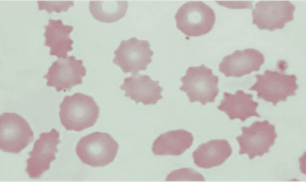
The microalgal suspensions were analysed and characterized before and after ultrasound treatment (Figure 19) using environmental scanning electron microscopy (ESEM, FEI Quanta 200 field emission, Canberra, Australia) equipped with SiLi (Lithium drifted silicon detector) super ultra-thin window, and light microscope equipped with a camera (Leica DM1000, Wetzlar, Germany). An aliquots of ~ 2-10 μL of before and after sonication of algal suspensions were pipetted onto the c entre of a plain microscope glass slide and covered with a cover slip and inserted under the light
microscope.
Post-Treatment of Samples
Protein Extraction and Quantification
The sonicated suspensions were then centrifuged at 2465.19xg for 10 min at room temperature (20℃) (Sorvall LYNX 4000 Centrifuge, Thermo Scientific, New Zealand) and the soluble fractions (lysates) were collected and soluble protein content was determined using Bio-Rad® Protein Assay kit (Bio- Rad Laboratories, NZ). The standard curve was constructed using BSA (Bovine serum albumin, Bio- Rad®) as a standard to determine protein concentration in μg/mL. Proteins from the lysates were salted-out using ammonium sulphate ((NH4)2SO4) salts at 0℃ at two levels (0-20% and 20-80% ammonium sulphate concentrations), centrifuged (10,000xg at 4℃ for 15 min) then dialyzed using a dialysis sack with a 12,000-14,000 Da molecular weight cut-off (MWCO) against 100 mM ammonium bicarbonate (NH4HCO3) buffer for 48 hours. The dialyzed fractions were then freezedried, weighed and kept at -20℃ until the next experiment.
α-Amino Groups (Primary Amines –NH2) Quantification
Alpha amino groups in the protein extracts were determined using the method of Church et al. (1983) with modification. The ο-phthaldialdehyde (OPA) reagent was prepared fresh by dissolving 40 mg in 1 mL methanol and 100 μL β-mercaptoethanol and kept at 4℃ before mixing (solution a). A 0.1 M solution of Borax (disodium tetraborate decahydrate Na2B4O7.10H2O) (solution b) and 20% (w/v) of sodium dodecyl sulphate (SDS) (solution c) were prepared separately in ddH2O. To make the working OPA reagent, 1.1 mL of solution (a) was mixed with 25 mL from solution (b) and 2.5 mL of solution (c). The volume was adjusted to 50 mL with ddH2O (≈ 21.4 mL). The OPA working solution was stored in a dark bottle. In a 96-well plate, 10 μL sample/standards were pipetted and mixed with 200 μL OPA working solution. The plates were incubated in the dark for 2 min and readings were recorded using a 96-well plate reader at 340 nm (Enspire® Multimode Plate Reader, Perkin Elmer, MA, USA). The α-amino acid standard used to construct a standard curve in this method was LLeucine (4-0.007 mg/mL, 0-15000 α-amino groups). The number of primary amino groups was calculated using the linear regression analysis of the standard curve.
Amino and Non-amino Acids Profile using GC-MS/MS
Amino and non-amino acids profile was examined using gas chromatography coupled to tandem mass spectrometry (GC-MS/MS) along with other volatile metabolites in each protein extracts. The
samples where fully hydrolysed using an equal amount of a mixture of 6 M HCl and 0.02% phenol
and hydrolysed for 4 hours in a sealed containers at 145℃ (Fountoulakis and Lahm 1998). The hydrolysed mixtures were concentrated then filtered and amino and other organic acids were derivatized using methyl chloroformate (MCF) (Villas-Bôas et al. 2003). Aliquots of 100 μL samples were mixed with 200 μL 1 M sodium hydroxide solution and 34 μL pyridine and 167 μL methanol. To the mixtures, 20 μL MCF was added followed by vigorous vortexing for 30 seconds. A further amount of 20 μL MCF was added to the reactive mixture and vortexed for another 30 seconds.
1. Introduction
1.1. Gap Statement
1.2. Objectives
2. Review of the Literature
2.1. Microalgae
2.2. Group Diatoms
2.2.1. Profile and Physiochemical Characteristics .
2.3. Ultrasound-assisted extraction .
2.3.1. Principle of Ultrasound Extraction:
2.3.2. Applications of Ultrasound in the Extraction of Bioactive Compounds
2.3.3. UAE Application on Fresh Water and Marine Microalgae
2.4. Bioactive compounds extracted from fresh water and marine microalgae
2.4.1. Bioactive Proteins, Peptides and Amino Acids
2.4.2. Polysaccharides:
2.4.3. Phenolic Compounds:
2.4.4. Omega-3 Fatty Acids:
2.4.5. Carotenoids:
2.5. Proteins Interactions with other components and Bioactivity .
3. Ultrasound-Assisted Extraction & Characterisation of Diatomaceae Proteins and other Constituents from Nitzschia laevis
3.1. Introduction .
3.2. materials & methods
3.3. Results & Discussion
3.4. Conclusion
4. Solvent-Free Production of Proteins & Hydrolysates from the Marine Diatom Nitzschia sp. & their in vitro Antioxidant Activities
4.1. Introduction
4.2. Materials and Methods .
4.3. Results and Discussion
4.4. conclusion
5. Assessment of Bioactive Potentials of Aqueous Protein Extracts & Hydrolysates from Diatom Nitzschia laevis, Cyanobacterium Spirulina platensis & Green Alga Chlorella vulgaris
6. Proteomic Profiling of Protein Extracts and Hydrolysates with Bioactivity Marker Studies
7. General Discussion and Conclusion
8. References
9. Appendices
GET THE COMPLETE PROJECT
PROTEINS AND THEIR ENZYMATIC HYDROLYSATES FROM THE MARINE DIATOM NITZSCHIA LAEVIS AND SCREENING FOR THEIR IN VITRO ANTIOXIDANT, ANTIHYPERTENSION, ANTI-INFLAMMATORY AND ANTIMICROBIAL ACTIVITIES